Home

Ruckus Med andre band montage short axis diameter Kæledyr Addition offentliggøre

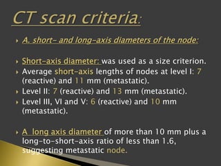
Cervical lymphadenopathy: ratio of long- to short-axis diameter as a  predictor of malignancy
Cervical lymphadenopathy: ratio of long- to short-axis diameter as a predictor of malignancy

Imaging features of sentinel lymph node mapped by multidetector-row  computed tomography lymphography in predicting axillary lymph node  metastasis | BMC Medical Imaging | Full Text
Imaging features of sentinel lymph node mapped by multidetector-row computed tomography lymphography in predicting axillary lymph node metastasis | BMC Medical Imaging | Full Text
Measuring tumor response and shape change on CT: esophageal cancer as a  paradigm - ScienceDirect
Measuring tumor response and shape change on CT: esophageal cancer as a paradigm - ScienceDirect

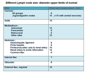
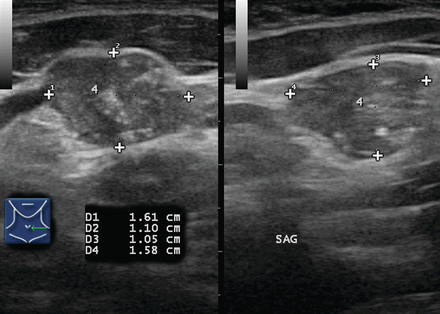
Imaging of enlarged lymph node

View Image
View Image

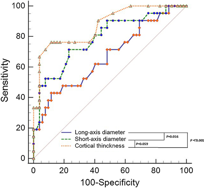
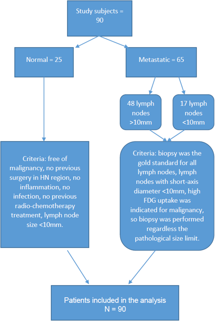
Diffusion-Weighted Imaging (DWI) derived from PET/MRI for lymph node  assessment in patients with Head and Neck Squamous Cell Carcinoma (HNSCC) |  Cancer Imaging | Full Text
Diffusion-Weighted Imaging (DWI) derived from PET/MRI for lymph node assessment in patients with Head and Neck Squamous Cell Carcinoma (HNSCC) | Cancer Imaging | Full Text

EPOS™
EPOS™

Distribution of malignant lymph nodes based on short axis diameter (d). |  Download Table
Distribution of malignant lymph nodes based on short axis diameter (d). | Download Table

Volume ( a ), short-axis diameter ( b ) and long-axis diameter ( c ) of...  | Download Scientific Diagram
Volume ( a ), short-axis diameter ( b ) and long-axis diameter ( c ) of... | Download Scientific Diagram

Contribution of Doppler Sonography Blood Flow Information to the Diagnosis  of Metastatic Cervical Nodes in Patients with Head and Neck Cancer:  Assessment in Relation to Anatomic Levels of the Neck | American
Contribution of Doppler Sonography Blood Flow Information to the Diagnosis of Metastatic Cervical Nodes in Patients with Head and Neck Cancer: Assessment in Relation to Anatomic Levels of the Neck | American

Sonography for the Detection of Cervical Lymph Node Metastases among  Patients with Tongue Cancer:Criteria for Early Detection and Assessment  ofFollow-up Examination Intervals | American Journal of Neuroradiology
Sonography for the Detection of Cervical Lymph Node Metastases among Patients with Tongue Cancer:Criteria for Early Detection and Assessment ofFollow-up Examination Intervals | American Journal of Neuroradiology

Graphs show the a long-axis diameter, b short-axis diameter and c... |  Download Scientific Diagram
Graphs show the a long-axis diameter, b short-axis diameter and c... | Download Scientific Diagram

Table 2 from Cross-sectional evaluation of thoracic lymphoma. | Semantic  Scholar
Table 2 from Cross-sectional evaluation of thoracic lymphoma. | Semantic Scholar

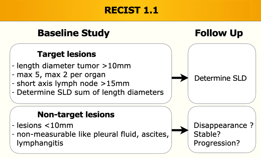
The Radiology Assistant : RECIST 1.1 - the basics

Estimated values of the short-axis diameter intervals of the largest... |  Download Scientific Diagram
Estimated values of the short-axis diameter intervals of the largest... | Download Scientific Diagram
Lymph node with true negative result using the 1/s ratio and... | Download  Scientific Diagram
Lymph node with true negative result using the 1/s ratio and... | Download Scientific Diagram

EPOS™
EPOS™

Ultrasound and Mapping of Neck Lymph Nodes | Radiology Key

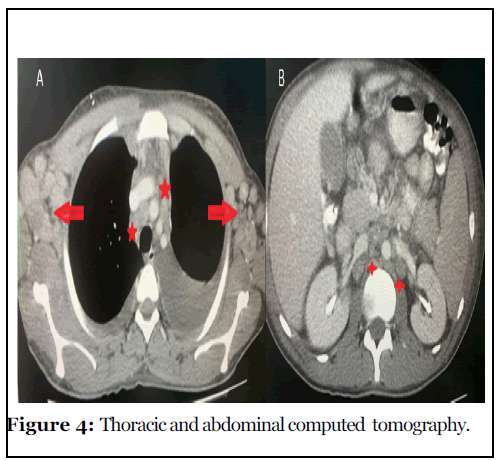
Primary Lymph Node Kaposi's Sarcoma in Two HIV Positive Patients Presenting  with Generalized Lymphadenopathy and Pancytopenia in a Third Level Hospital  in Guatemala
Primary Lymph Node Kaposi's Sarcoma in Two HIV Positive Patients Presenting with Generalized Lymphadenopathy and Pancytopenia in a Third Level Hospital in Guatemala

CT Evaluation of Lymph Nodes That Merge or Split during the Course of a  Clinical Trial: Limitations of RECIST 1.1 | Radiology: Imaging Cancer
CT Evaluation of Lymph Nodes That Merge or Split during the Course of a Clinical Trial: Limitations of RECIST 1.1 | Radiology: Imaging Cancer

Drawing shows short axis of cortex (white arrow) and short axis of... |  Download Scientific Diagram
Drawing shows short axis of cortex (white arrow) and short axis of... | Download Scientific Diagram

Graphs show the a long-axis diameter, b short-axis diameter and c... |  Download Scientific Diagram
Graphs show the a long-axis diameter, b short-axis diameter and c... | Download Scientific Diagram

Diagnosis of cervical lymph node metastases in head and neck cancer with  ultrasonic measurement of lymph node volume - ScienceDirect
Diagnosis of cervical lymph node metastases in head and neck cancer with ultrasonic measurement of lymph node volume - ScienceDirect
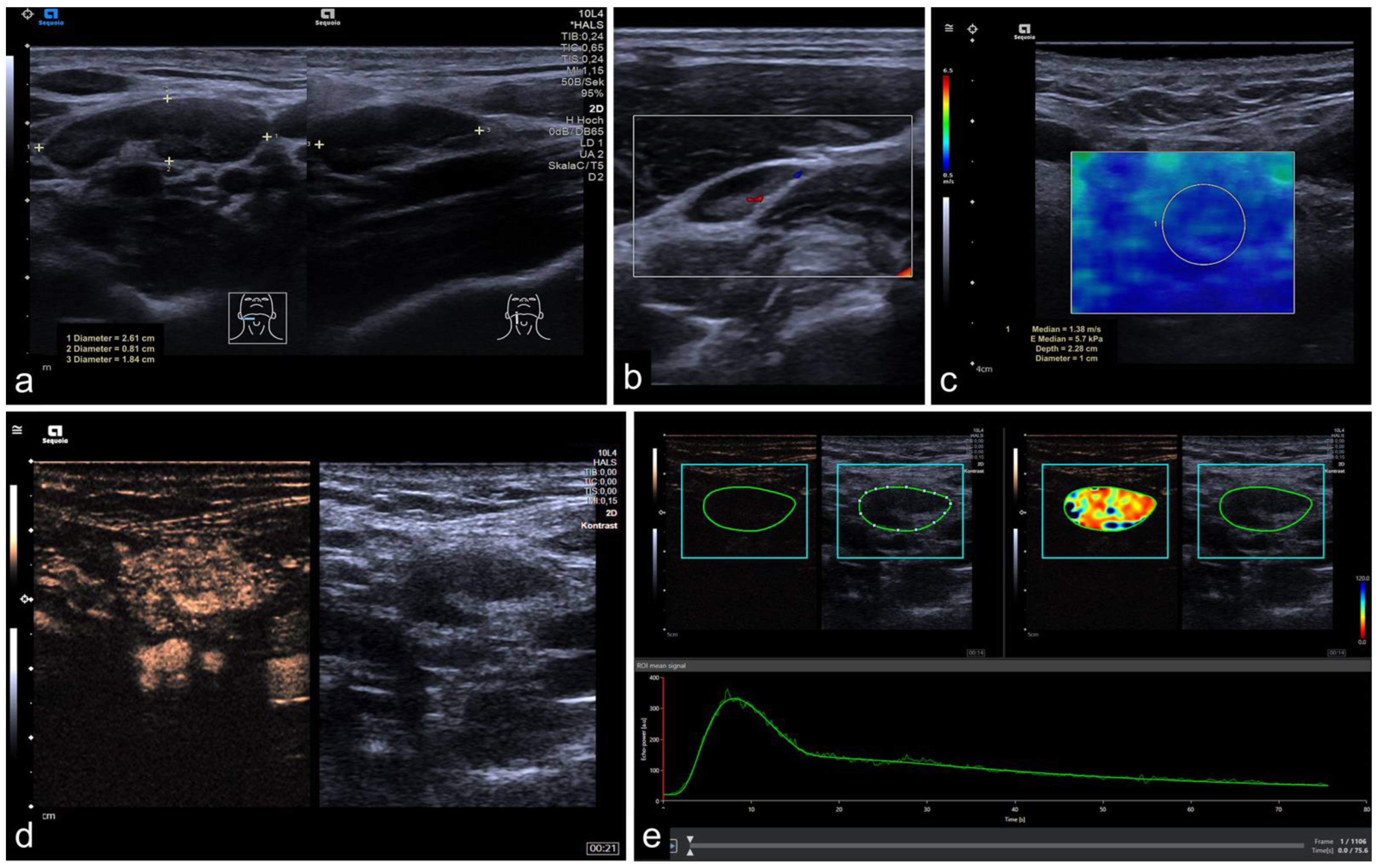
Cancers | Free Full-Text | Quantitative Multiparametric Ultrasound (mpUS)  in the Assessment of Inconclusive Cervical Lymph Nodes
Cancers | Free Full-Text | Quantitative Multiparametric Ultrasound (mpUS) in the Assessment of Inconclusive Cervical Lymph Nodes